 Table 1-2
Chapter 1 and 2
1. Immunity
@ protective methods;
- physical barriers
- phagocyte cells in blood and tissues
- lymphocytes
- various blood-borne molecules
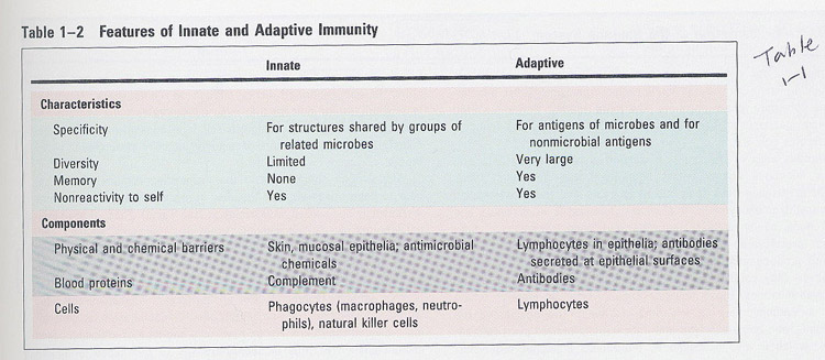
* Table 1-2
① innnate immunity
-> are present prior to exposure
-> not enhanced by exposure
-> phagocytes (Macrophages , NK), complements
② adaptive immunity
-> induced by exposure
-> specific for distinct macromolecules
-> remember (memory)
-> amplify the innate immunity
-> lymphocytes, antibody
Table 1-2
2. Types of adaptive immunity (Fig 1-2)
㉮ humoral: transfer by cell-free portion of blood (serum)
㉯ cell-mediated:
transfer by cells from immmunized individuals
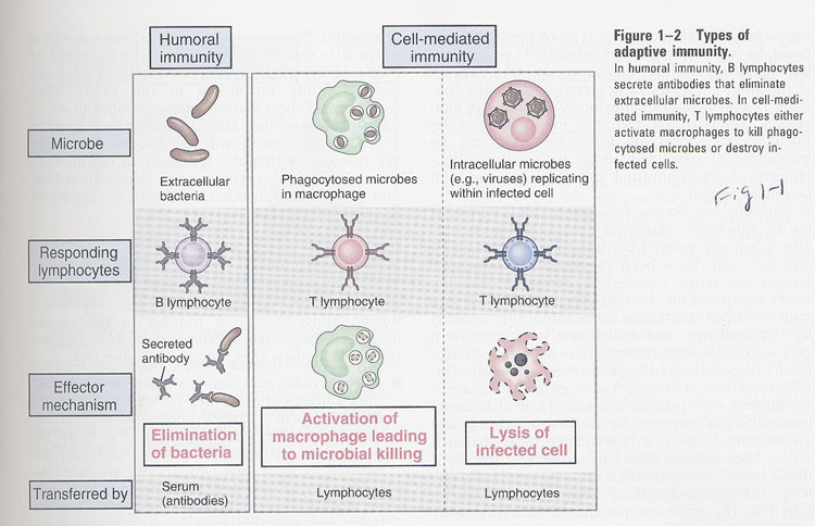 Fig 1-2
3. Features of Adaptive Immune response
(table 1-3)
1) specificity; for antigens, depend on epitope (determinant)
2) diversity; discrimination of 109 Ags
3) memory (Fig 1-4); 2' response is faster and more effective
4) self-limitation; IR is wane with time ---> basal state ; homeostasis
5) discrimination between self and non-self; tolerance/autoimmune disease
6) specialization; generate responses that are optimal for defense against different types of microbes
/AllImagesFromCellular_and_Molecular_Immunology_7E-1/S9781437715286-001-f004.jpg)
4. Phase of adaptive immune response
(Fig 1-6)
① recognition (cognitive); Ag recognition
② activation; proliferation (clonal expansion) and differentiation
③ effector; Ag elimination
④ homeostasis
⑤ memory
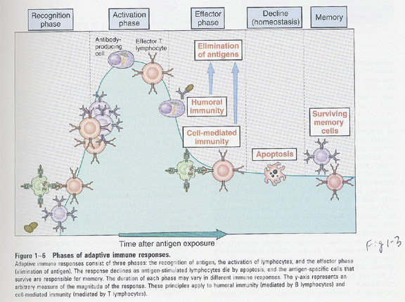 Fig 1-6
5. How a diverse repertoire can be generated ?
① instruction theory
-> by changing the conformation
of Ag binding region
② clonal selection theory (Fig 1-7)
-> each clone arise from
a single precursor
-> select and activate a
specific pre-existing clone
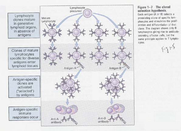 Fig 1-7
6. Cells of the immune system
(1) lymphocytes
① B lymphocytes; bird's bursa, bone marrow
② T lymphocytes; bone marrow -> thymus
-- morphology (Fig 3-4)
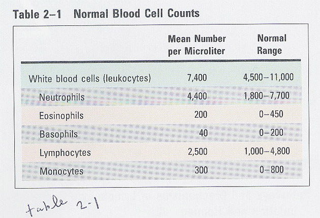 Table 3-1
(2) kinds of lymphocytes
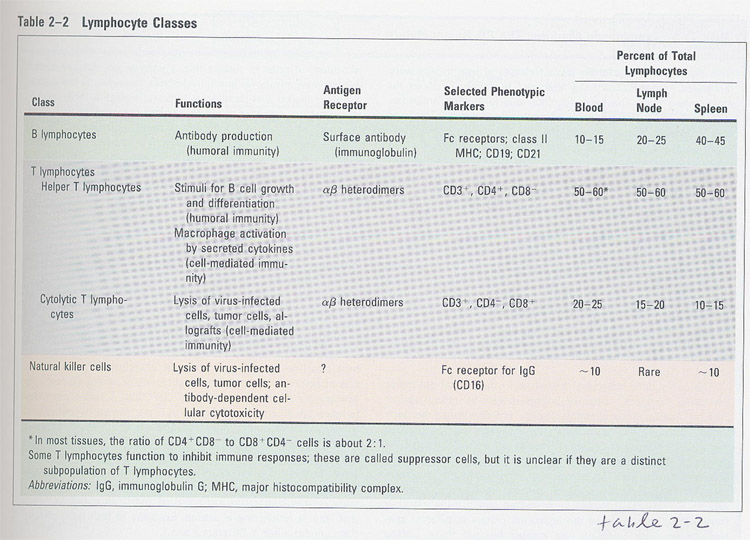 Table
3-2
Table
3-2
(3) Development of lymphocytes
- generative organs; lymphocytes arised and matured
- peripheral organs; mature lymphocytes recognize Ags
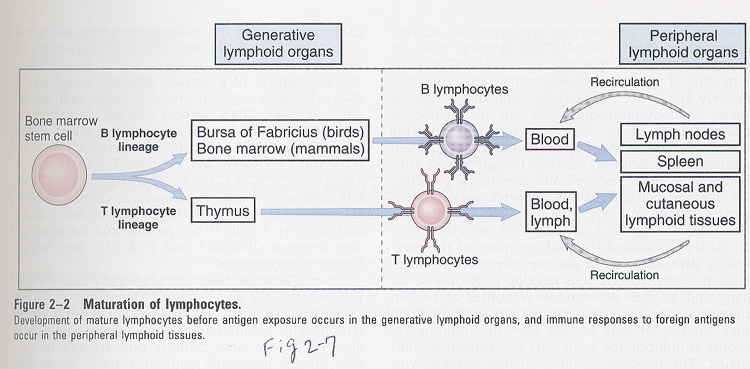 (Fig 3-2)
(4) Activation of lymphocytes
--- the phase of lymphocyte activation;
new protein synthesis (ex; cytokine) --> proliferation --> differentiation --> homeostasis
- morphological changes in activation
larger: lymphoblast, G0->G1
differentiated CTL; more granules
plasma cells ; -> don't circulate in blood, no mitotic
division
->
only in lymphoid organs
/AllImagesFromCellular_and_Molecular_Immunology_7E-1/S9781437715286-002-f008.jpg)
memory
lymphocytes; -> survive for a long time
->
surface marker
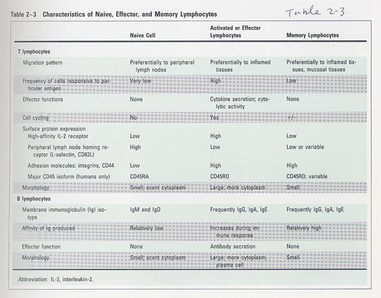 (table 3-3)
Ag ----->
 --------> normal
immune response
--------> normal
immune response
Ag -------> irradiation
-----------> X
lymphocytes -------> ----------->
normal immune response
(2) Antigen-presenting cells
1) Mononuclear Phagocytes
- development
/AllImagesFromCellular_and_Molecular_Immunology_7E-1/S9781437715286-002-f002.jpg)
-- morphology
/AllImagesFromCellular_and_Molecular_Immunology_7E-1/S9781437715286-002-f003.jpg)
- functions;
1) in innate immunity
-> phagocytosis
-> produce cytokines
and recruit infammatory cells
-> produce growth
factors and help the repair of injured tissues
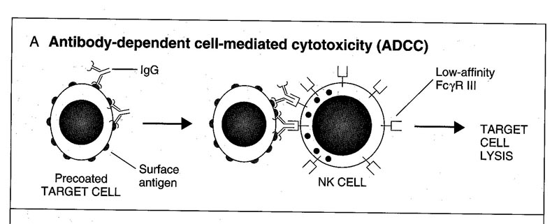
2) in adaptive immunity (ADCC)
-> APC
-> Fc receptor/
complement receptor and phagocytose
2) Dendritic cells
- spine-like projections
- types; ① interdigitating
dendritic cells
->
from bone marrow
->
interstitium of most organs
->
a good APC
->
related to phagocytes
②
follicular dendritic cells (FDC)
->
in germinal center of lymph node, spleen
->
not from bone marrow
- plasmacytoid DC; early cellular responders to viral infection
produce type 1 IFN
7. Lymphoid tissues
1) bone marrow
- hematopoiesis ; generation of all blood cells
/AllImagesFromCellular_and_Molecular_Immunology_7E-1/S9781437715286-002-f009.jpg)
- hematopoietic cytokines; ① by stromal
cells
②
macrophages
③
T cells stimulated by Ags
2) thymus
morphology
- cortex; dense T cells (CD4/CD8 double negative)
- Hassall's corpuscles; epithelial cells
/AllImagesFromCellular_and_Molecular_Immunology_7E-1/S9781437715286-002-f010.jpg)
/AllImagesFromCellular_and_Molecular_Immunology_7E-1/S9781437715286-002-f011.jpg)
3) lymph nodes
- throughout the body
- primary follicles (w/o germinal center) -> resting B not stimulated
by Ags
secondary follicles (w "
) -> B cells stimulated by Ags
- T cell zone (paracortex); CD4 positive
- Ag's pathway; by lymphatic
- medulla; macrophages, dendritic cells, plasma cells
- shape is flexible depend on Ag exposure; ex) germinal center within
1 week
Fig/AllImagesFromCellular_and_Molecular_Immunology_7E-1/S9781437715286-002-f012.jpg)
/AllImagesFromCellular_and_Molecular_Immunology_7E-1/S9781437715286-002-f013.jpg)
4) spleen
①
periarteriolar lymphoid sheath; T cells (2/3; CD4, 1/3; CD8)
white ② marginal zone; both B and T cells, macrophages
pulp ③ germinal center; B cells
④ red pulp; erythrocyte, macrophages, dendritic cells, plasma cells
/AllImagesFromCellular_and_Molecular_Immunology_7E-1/S9781437715286-002-f015.jpg)
8. Cutaneous immune system
-- skin, lymphocytes and APCs
Fig/Fig2-14%20copy.jpg) (Fig 3-12)
(Fig 3-12)
9. Mucosal immune system
-- in gastrointestinal and respiratory tracts
-- cells; lymphocytes and APCs
-- each region has distinct lymphocytes; ex) intraepithelial (CD8+), lamina propia (CD4+), Peyer's patch (CD4+)
-- M(membranous) cells --> delivering of antigen to Peyer,s patch
Fig/Fig2-15.jpg) Fig. 3-13
Fig. 3-13
10. Lymphocyte homing
/AllImagesFromCellular_and_Molecular_Immunology_7E-1/S9781437715286-003-f006.jpg)
/AllImagesFromCellular_and_Molecular_Immunology_7E-1/S9781437715286-003-f003.jpg)
Chapter 5 Antibodies
and Antigens
1. Antibody
① a related glycoprotein, no enzyme
② Ag binding (non-covalent), specific
③ BCR
④ other Ag recognizing molecules (MHC, TCR)
/AllImagesFromCellular_and_Molecular_Immunology_7E-1/S9781437715286-005-f001.jpg)
2. Natural distribution
of Ab
-> fluid portion of blood
-> cytoplasmic membrane-bound compartments (ER, Golgi)
-> surface of B cells and other cells (phagocytes, NK, mast cells)
-> interstitial fluid of tissues
-> secretory fluids (mucus, milk)
3. Structure of Ab
① all Abs are similar in overall structure
② 2 heavy + 2 light
CDR (complementarity
determining region, ~10 Aas); a hypervariable region
/AllImagesFromCellular_and_Molecular_Immunology_7E-1/S9781437715286-005-f005.jpg)
FW (Frame work region)
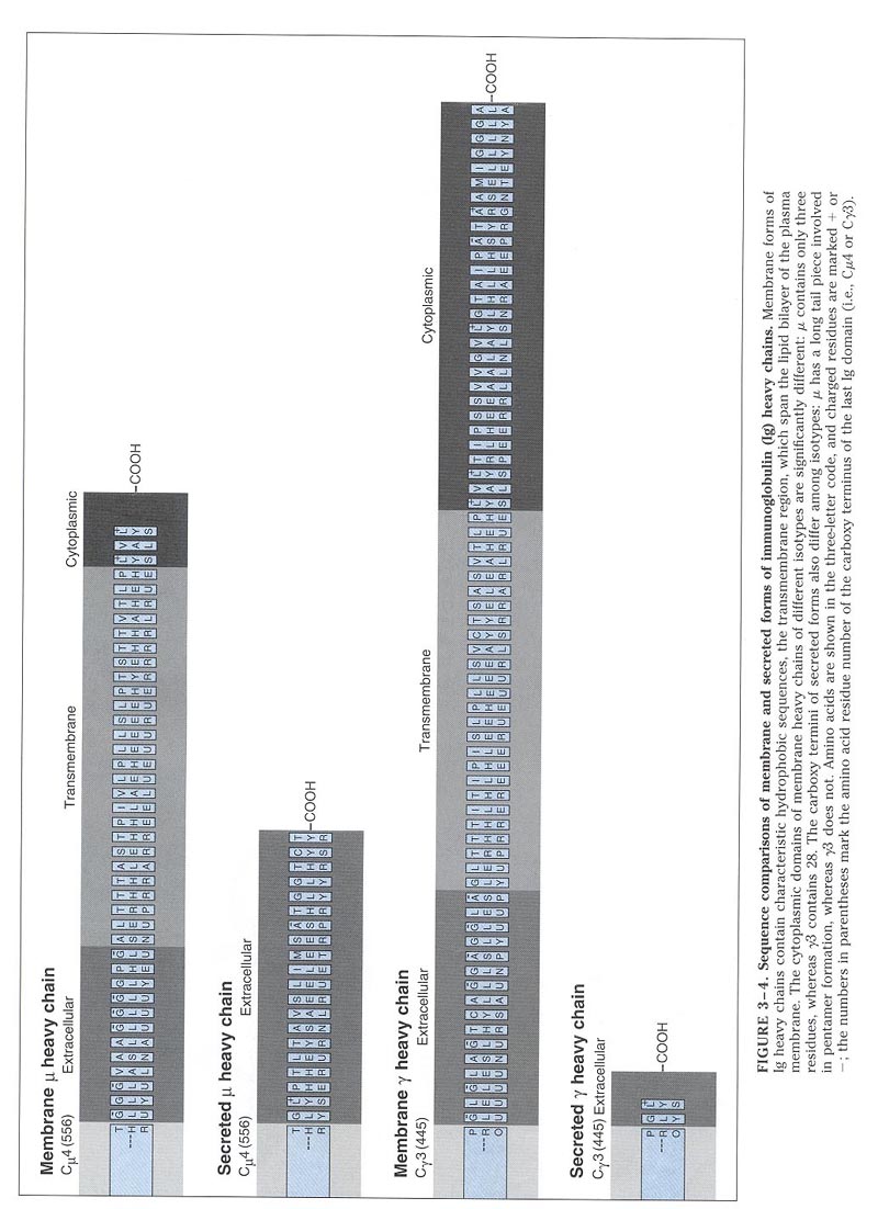
1) H chain
-> CDR3 is most variable
-> hinge region; steric effect
-> two forms (Fig 4-7); membrane and secreted form
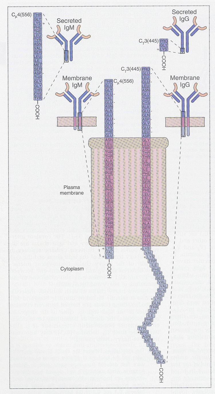
-> tail pieces (Table 4-2);
extended nonglobular seq. in secreted form
intermolecular
interactions (multimeric form)
IgM(5),
IgA(2)
-> J chain; S-S bonding between tail pieces -> stabilization
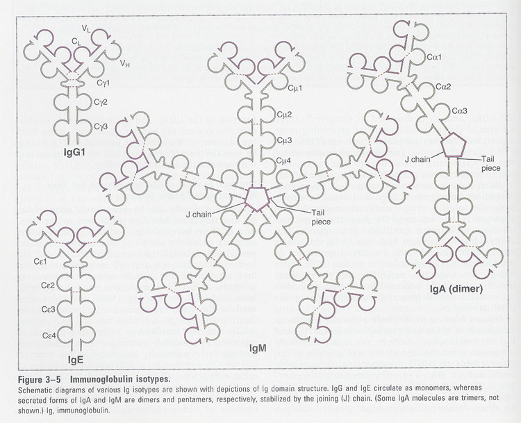 Table 4-2
2) L chain
-> kappa and
lamda , (human 50:50; mouse 10 times in kappa )
-> CDR3 is most variable
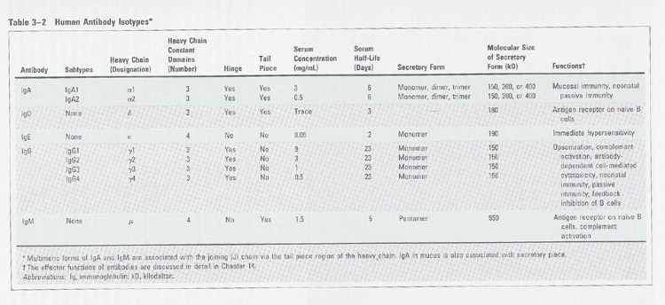
③ isotype (table 4-2); A, D, E, G, M -> different effector functions
④ Fab and Fc
--- Fab; Ag-binding
--- Fc; crystalline
--- Fab'; include
hinge region
/AllImagesFromCellular_and_Molecular_Immunology_7E-1/S9781437715286-005-f003.jpg)
4. Maturation of B lymphocytes
/AllImagesFromCellular_and_Molecular_Immunology_7E-1/S9781437715286-005-f010.jpg)
1. pre-B lymphocytes; - only cytoplasmic
μ heavy chain
(associated
with surrogate light chain (Vpre-B
and λ5): prevent
the degradation of newly synthesized μchain, promote the Igs on
the cell surface)
-
bone marrow and fetal liver
-
nonresponse to antigen
2. immature B lymphocytes; - synthesis
of light chain
-
membrane IgM
-
not proliferate and differentiate by Ag
(self
Ag - nonresponse; tolerance)
-
bone marrow
3. mature B cells; - coexpress the
IgM/IgD: same antigen specificity
-
response to Ag, but can mature without Ag, however they will die (half life=3-4days)
-
peripheral blood and lymphoid tissues
4. activated B cells; - Ag stimulated,
and proliferate and differentiate (sIg>mIg)
-
some: heavy chain class switching
others:
keep the membrane form of Ig (memory B cells)
5. antibody secreting cells; - plasma cells
5. antigenic determinant (= epitope)
-- features of biologic antigens
1) conformational determinant
2) linear determinant
3) neoantigenic determinant
/AllImagesFromCellular_and_Molecular_Immunology_7E-1/S9781437715286-005-f012.jpg)
-- structural and chemical basis of antigen binding
/AllImagesFromCellular_and_Molecular_Immunology_7E-1/S9781437715286-005-f013.jpg)
6. changes of antibody structure after IR
/AllImagesFromCellular_and_Molecular_Immunology_7E-1/S9781437715286-005-f015.jpg)
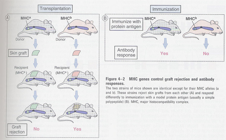
Chapter 6 MHC (major histocompatibility complex) and Antigen processing
General features;
(1) major determinant of graft rejection (Fig 5-2)
(2) highly polymorphic, in various cells
(3) membrane-associated and not secreted
(4) involved in T cell development
1. kinds of MHC
1. Class I MHC molecules
/AllImagesFromCellular_and_Molecular_Immunology_7E-1/S9781437715286-006-f010.jpg)
- heterodimer ( α + β2 -microglobulin; non-covalent association)
a) α chain; by MHC genome, and highly polymorphic
heavy
chain
5
domains (3 extracellular: α1, α2, α 3; transmembrane; cytoplasmic)
N-linked
glycosylation: human (1), mouse (2)
b) β2 microglobulin; by non-MHC genome
non-attachment to the cells
2. Class II MHC molecules
/AllImagesFromCellular_and_Molecular_Immunology_7E-1/S9781437715286-006-f012.jpg)
- heterodimer ( α+ β)
a) in overall structure, α and β chain are similar
b) α> β ; due to the more glycosylation
c) α and β chain are encoded by MHC genome
d) α and β chain are polymorphic
2. Structure of MHC loci
General features:
A. in human and mouse
/AllImagesFromCellular_and_Molecular_Immunology_7E-1/S9781437715286-006-f007.jpg)
/AllImagesFromCellular_and_Molecular_Immunology_7E-1/S9781437715286-006-f008.jpg)
(1) ~3500 kb DNA fragment
(2) in class II; 2-3 funcional β chains and 1 functional α chain ->
more than two alleles
pseudo
genes (DZ, DO, DX)
(3) in class III; complement (C2, C4)
(4) cytokines; TNF and LT
(5) class I; class I - like genes
(some is pseudo genes; some is non-polymorphic proteins associated with β2-microglobulin),
functions are unknown.
called
as class 1B proteins
ex:
① Tla (mouse thymocytes)
②
Fc receptor-like protein; IgG transport across epithelial barrier
③
CD1; presenting of non-peptide antigens to unusual T cell populations
④
HLA-H; iron metabolism
possible role; a repository for nucleic acid used for polymorphic sequence of class
I (gene conversion -> new alleles)
B. in mouse
(1) smaller region
(2) class I - class II - class I
3. Expression of MHC molecules
1. regulatory sequences
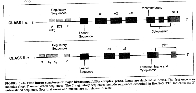
- target for DNA binding proteins
- in class I; for cytokines
(ex; interferon); A (enhancer A), ICS (interferon consensus sequence), B (enhancer
B)
in class II; S, X, Y box necessary for gene expression
2. expression
- class I : CTL -- most cells
- class II : T helper cells -- a few cells
/AllImagesFromCellular_and_Molecular_Immunology_7E-1/S9781437715286-006-f014.jpg)
3. regulation of expression
- 3 important features
(1) transcriptional rate
(2) coordinate regulation of transcription and expression
class I; between α and β2-microglobulin
class II; between γ and α,β
(3) cytokine modulation
class I; induced by γ-IFN, α,β-IFN, TNF, LT
class II; induced by γ-IFN, IL-4
Fig/Fig4-11.jpg) Fig 5-10
Fig 5-10
4. Biosynthesis of MHC
- ribosome -> ER -> Golgi -> plasma membrane
(1) in class I
a) within ER, α and β2-microglobulin are associated
b) role of β2-microglobulin;
transport
evidence: Daudi B cells deficient
β2-microglobulin are transfected with β2-microglobulin,
and check the expression
or
using cell fusion with β2-microglobulin expressing cells
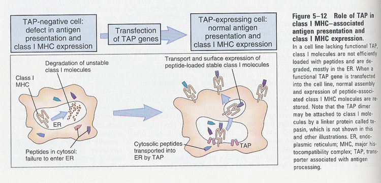
(i) class I MHC-restricted CD8+ T cell 에서의 antigen presentation
1) 세포질내에서 단백질 항원의 합성 (예를들어 바이러스 단백질) 이나 세포질내로
단백질 항원의 전달
2) ubiquitination을 통한 proteasome에 의해서 peptides로 분해
3) TAP (transporter associated with antigen presentation) transport에 antigenic fragment (즉 분해되어진
peptides)의 결합 --> class 1 assembly and surface expression
 Fig
6-14
Fig
6-14
4) ER에서 이미 생성되어 있는 class I MHC와 상호결합
5) Golgi를 통하고 궁극적으로는 secretory vesicle의 세포막 융합에 의하여
발현되어지는 것이다.
/AllImagesFromCellular_and_Molecular_Immunology_7E-2/S9781437715286-006-f016.jpg)
(2) in class II
a) within ER, α and βchains
are coordinately associated
b) γ(invariant) chain:
- ~30 kDa, Ig superfamily, Type
II protein
- non-polymorphic, and not encoded
by MHC loci
- associated with a recently
synthesized class II
- separated from before class
II expresses on surface
functions:
1) direct the trafficking
2) prevent association of endogenously
synthesized peptides with class II
(ii) class II MHC-restricted CD4+ T cell 에서의 antigen presentation
/AllImagesFromCellular_and_Molecular_Immunology_7E-2/S9781437715286-006-f017.jpg)
- antigenic fragment 생성은 세포내 기관인 lysosome, endosome에서 일어난다.
1) 항원으로써의 역할을 수행하는 단백질의 세포내로 흡입 (endocytosis)
2) endosome, lysosome에서 단백질 항원의 분해, peptide 생성
3) 한편 ER에서는 class II MHC가 생성되어지나 항원과의 상호결합은 없는
상태를 유지함, 이때 Ii (invariant chain)이 antigenic fragment 결합부위를 보호하고 있다.
4) Golgi를 통과한 class II MHC을 보유하고 있는 secretory vesicle 와 2)의 endosome의
융합, 따라서 Ii chain은 class II MHC으로부터 분리되고 대신에 endosome내의 peptide가
결합되어 진다.
** CLIP (class II-associated invariant chain peptide)
HLA-DM (calss II like molecule) ---> binds the CLIP
/AllImagesFromCellular_and_Molecular_Immunology_7E-2/S9781437715286-006-f019.jpg)
5. MHC restriction
--> Fig
6-15 ; self-MHC and foreign Ags
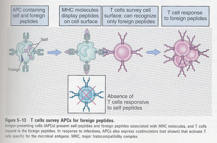
-- MHC restriction of CTL
/AllImagesFromCellular_and_Molecular_Immunology_7E-1/S9781437715286-006-f006.jpg)
--> -
class I : CTL (CD8 positive)
- class II : T helper cells (CD4 positive)
/AllImagesFromCellular_and_Molecular_Immunology_7E-1/S9781437715286-006-f015.jpg)
6. Antigen processing
--> antigen features; recognized by T cells
Fig/table5-1.jpg) Table 6-1
Table 6-1
--> Requirement of APC, not antigen itself, for T cell activation
Fig/Fig5-2.jpg) Fig 6-2
Fig 6-2
--> Functions of different APCs
/AllImagesFromCellular_and_Molecular_Immunology_7E-1/S9781437715286-006-f002.jpg)
/AllImagesFromCellular_and_Molecular_Immunology_7E-2/S9781437715286-006-f021.jpg)
--> Comparison of MHCI- and II-derived antigen presentation
Fig/table5-4.jpg) Table 6-3
Table 6-3
--> Antigen processing requires time and cellular metabolism (Fig 6-10)
Fig/Fig5-11.jpg)
Chapter 7 Antigen Receptors and Signal Transduction
TCR and accessory molecules
/AllImagesFromCellular_and_Molecular_Immunology_7E-2/S9781437715286-007-f009.jpg)
-- Comparison of TCR and BCR signaling
Fig/Fig6-2.jpg) Fig 7-2
Fig 7-2
** B cell antigen receptor (BCR) **
/AllImagesFromCellular_and_Molecular_Immunology_7E-2/S9781437715286-007-f005.jpg)
-- TCR and BCR
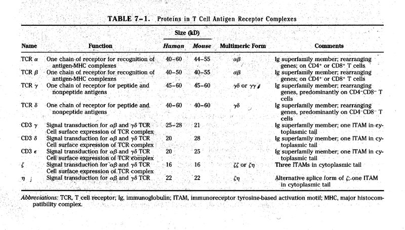
Fig/table6-2.jpg) table 7-1
table 7-1
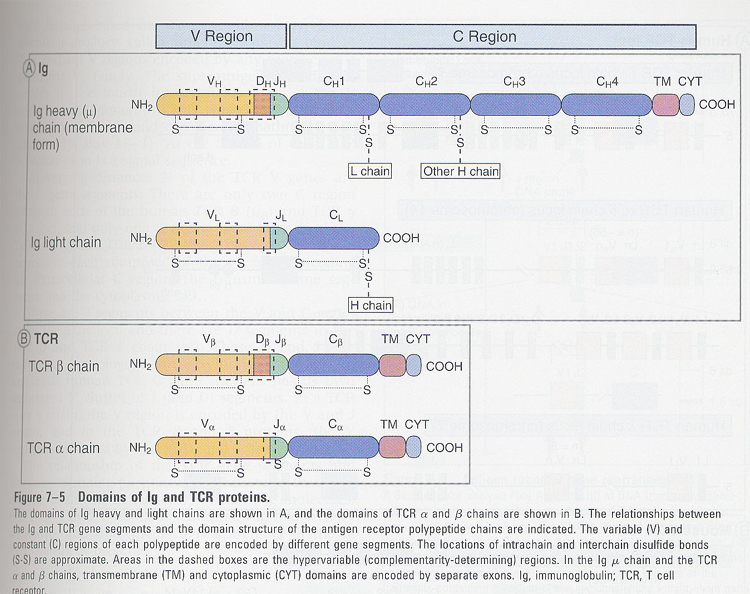
- - TCR
/AllImagesFromCellular_and_Molecular_Immunology_7E-2/S9781437715286-007-f006.jpg)
1) αβ TCR
-> S-S bond
-> α chain; acidic,
β chain; basic and charged
-> each C region; functional domains
-> TM region; + charged (Lys, Arg)
-> cytoplasmic tail; 5-12 Aas
- differences
between α, βchains and Ig
-> no isotype switching
-> don't know the effector functions
2) γδTCR
- in small subset of peripheral T cells
in immature thymocytes and bowel
mucosal
- features;
1) has a CD3 complex
2) β = δ , α =
γ
3) S-S bond and non-covalent binding
4) γδrearrangement precedes those
of αβ
5) don't express CD4 or CD8
6) no MHC restriction, non-specific
cytolytic activity
7) what γδ TCR recognizes ?
-> MHC
I like proteins (CD1, Tla)
-> heat
shock proteins of Mycobacterium
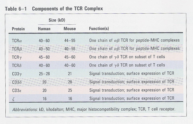
3) CD3 complex, table 6-1
/AllImagesFromCellular_and_Molecular_Immunology_7E-2/S9781437715286-007-f008.jpg)
1) require for surface expression
- evidence; ① coprecipitation
②
both endocytosis
③
reconstitution of mutant cell line (TCR-); (Fig 7-6)
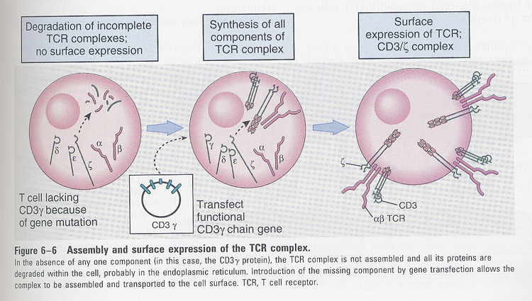 Fig
7-6
Fig
7-6
2) γδεζηchains
- ζη(10%), ζζ(90%)
- highly homologous,
gene duplication
- no polymorphism
- TM; Asp(-)
Accessory molecules of T cells
* Interaction between T cells and APC (Fig 7-7)
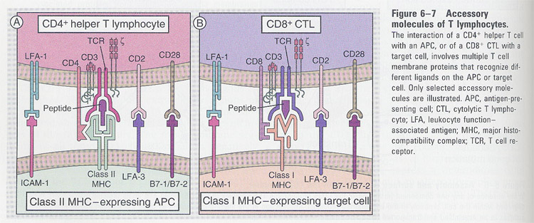 Fig 7-7
CD4 and CD8
Fig/Fig6-9.jpg) Fig 7-8
Fig 7-8
- MHC restriction
- Ig gene superfamily
- not related each other, but similar functions
- in peripheral T cells (CD4; 65%, CD8; 35%)
1) CD4
- as a monomer
- functions; ① cell-cell adhesion ② signaling
evidences;
CD4
![]()
fibroblast
fibroblast
![]()
![]()
+
MHC II expressing cells
![]()
![]()
![]()
no-binding
binding
(1)
mAB against CD4 --> stimulatory or inhibitory
(2) cytoplasmic tail --> phosphorylation
2) CD8
- CD8 α and β, heterodimer and homodimer (αα)
- functions; ① cell-cell adhesion ② signaling
evidences;
(1)
between fibroblast and MHC I expressing cells
(2)
mAB against CD8 --> stimulatory or inhibitory
(3)
cytoplasmic tail --> phosphorylation
Fig/Fig6-10.jpg)
Adhesion molecules; integrin
-- role of integrin in T cell response
Fig/Fig6-11.jpg)
Chapter 8 Lymphocyte maturation & Antigen receptor gene rearrangement
-- Stages of Lymphocyte maturation
/AllImagesFromCellular_and_Molecular_Immunology_7E-2/S9781437715286-008-f001.jpg)
-- checkpoints of lymphocyte maturation (Fig 8-3)
--> only
cytoplasmic μ heavy chain
(associated with
surrogate light chain (Vpre-B
and λ5): prevent
the degradation of newly synthesized μchain, promote the Igs on
the cell surface)
/AllImagesFromCellular_and_Molecular_Immunology_7E-2/S9781437715286-008-f003.jpg)
Sequence of events in thymocyte maturation (Fig 8-20)
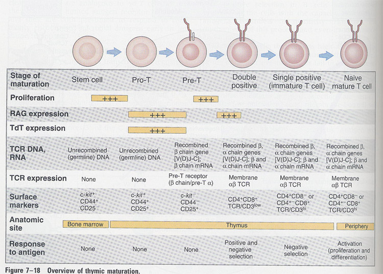 Fig 8-20
pre-Tα
chain (pTα)
- analogous to surrogate light chain ( λ5)
- 3 important functions
① recombination at α chain locus; allelic exclusion of β chain
② stimulation of proliferation
③ transition from double-negative to double-positive cells; induction
of CD4 and CD8
Ontogeny of T cells (Fig 8-21)
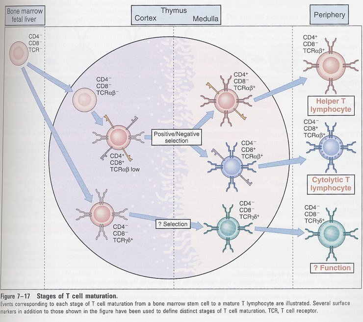
- in thymus
① MHC; by non-lymphoid cells
② thymic hormone; by epithelial cells
③ IL-7; by stromal cells
- positive and negative selection
1) positive selection -- TCR binds self-MHC (low
affinity) --> can survive
2) negative selection -- TCR binds self-Ag (high
affinity) --> can die (clonal deletion)
or
can be inactivated (clonal anergy)
positive & negative selection
/AllImagesFromCellular_and_Molecular_Immunology_7E-2/S9781437715286-008-f004.jpg)
positive and negative selection in T cells (Fig 8-24)
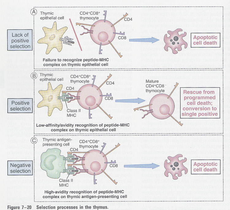 Fig 8-24
-- Role of peptide in positive selection (Fig 8-26)
Fig/Fig7-22.jpg) Fig 8-26
Fig 8-26
Formation of functional antigen receptor genes in B and T lymphocytes
1. Genomic organization of Ig
/AllImagesFromCellular_and_Molecular_Immunology_7E-2/S9781437715286-008-f005.jpg)
1. V region; 300bp,
1000 to 2000kb long
leader(=signal)
peptide is 90bp of 5' of each V region, hydrophobic
from
ribosome to lumen of ER
cleavage
before complete translation
duplication
of a single V gene
2. C region; tandem
array whose order is specific for each species
3. J region; 30-50bp long, 3'hypervariable region
4. D region; only in heavy chain, 3'hypervariable region
5. intron region; important role in the production of Ab
-
recognition seq for rearrangement, RNA splicing, regulation of transcription
2. Organization of
TCR
/AllImagesFromCellular_and_Molecular_Immunology_7E-2/S9781437715286-008-f007.jpg)
3. Antigen receptor gene rearrangement
/AllImagesFromCellular_and_Molecular_Immunology_7E-2/S9781437715286-008-f008.jpg)
4. Sequence of Ig gene rearrangement (somatic recombination)
/AllImagesFromCellular_and_Molecular_Immunology_7E-2/S9781437715286-008-f015.jpg)
H
chain:
1. DJ joining
-- no-effect on 5' D and 3' J region
-- before commitment of B precursors to B lineage cells
2. VDJ joining
-- only in cells committed to become B cells
-- not known whether all the C regions can be expressed in primary
transcript
L
chain:
1. VJ joining
5. Mechanism of Ig
gene rearrangement
1. recombinases;
- cell type specific (ex: B and T cells)
- function at early stage of B cell development (pre-B cells)
- multiple enzymatic activities (some are B cell specific, others
are not; exonuclease and ligase)
- RAG1/RAG2; genetic defect in SCID mice (no mature B and T cells)
** recognition sequence
;
- located in 3' V, 5' J, and both side of D region
- heptamer(CACAGTG)/nonamer(ACAAAAACC): conserved
12 and 23 spacers: non-conserved
/AllImagesFromCellular_and_Molecular_Immunology_7E-2/S9781437715286-008-f012.jpg)
2. accessibility of DNA to recombinase
- chromatin; open in early B cell stage
-> euchromatin (vs: heterochromatin)
3. programmed change or selection pressure by
environmental antigens
- in pre-B, proximal Vh region is preferred
- in mature B, no bias in Vh usage
6. Antibody diversity (table 8-1)
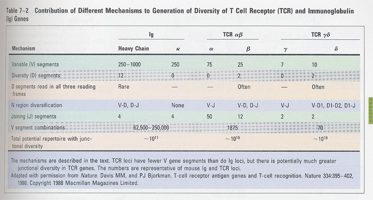
1. mutiple germline genes
- multiple germline V, D, J genes
2. combinational diversity: V X D X J
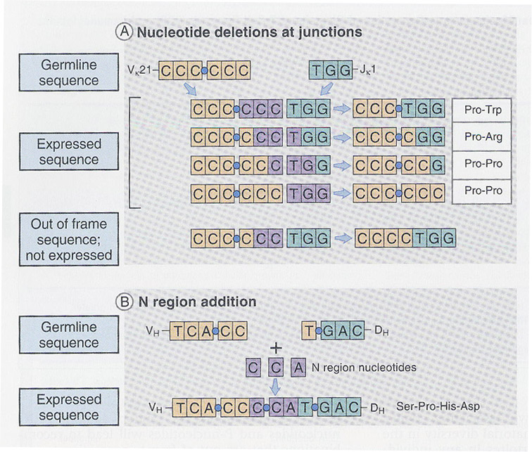
3. junctional diversity (Fig
8-13); more important in TCR diversity
(a) imprecise DNA rearrangement: functional, nonfunctional(occasional compensation by deletion
of 1 or 2 nt)
(b) N region diversification (addition) / P nucleotide addition
- N sequence is not in germline
- random process, by TdT(terminal deoxyribonucleotidyl
transferase)
- P nucleotide addition; by RAG1/2 and DNA polymerase
Fig/Fig7-11.jpg)
/AllImagesFromCellular_and_Molecular_Immunology_7E-2/S9781437715286-008-f013.jpg)
4. somatic mutations
5. combination between H and L chain
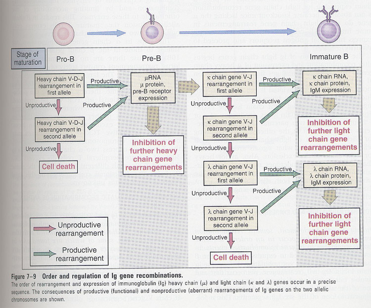
7. B cell maturation
/AllImagesFromCellular_and_Molecular_Immunology_7E-2/S9781437715286-008-f014.jpg)
** Roles of heavy chain
1. inhibition of rearrangement on other
chromosome (allelic exclusion)
-- nonproductive; formation of stop codon by deletion, mutation,
frame-shift
finally
die (no response to Ag)
2. stimulation of L chain rearrangement
-- k, and λ chian rearrangement
** role of k light chain; allelic exclusion and light
chain isotype exclusion
(a) only k-producing B cells: lamda gene is a germline or
unarranged
(b) only λ-producing B cells: k gene is deleted or aberrantly
recombined
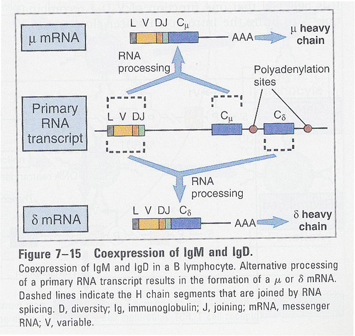
** Coexpression
of IgM and IgD
- in mature B cells
- same antigen specificity - same V region
- alternative RNA splicing
8. T cell maturation
/AllImagesFromCellular_and_Molecular_Immunology_7E-2/S9781437715286-008-f019.jpg)
-- T cell maturation in thymus (Fig 8-21)
-- TCR recombination; similar to Ig
-- positive and negative selection in thymus
Chapter 9 Activation of T lymphocytes
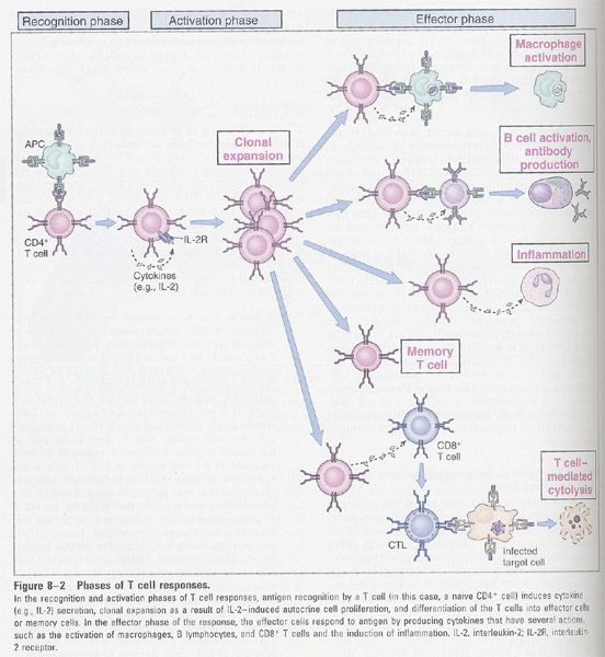
-- How T cells are activated ? (Fig 9-1)
Fig/Fig8-1.jpg)
(a) effector function
- T helper cells secrete cytokines
--> (Fig 9-2)
- CTL lyse the Ag-bearing target cells, and secrete
cytokines
(b) proliferation
- autocrine pathway (by IL-2)
-> clonal expansion and Ag-specific
memory T cells
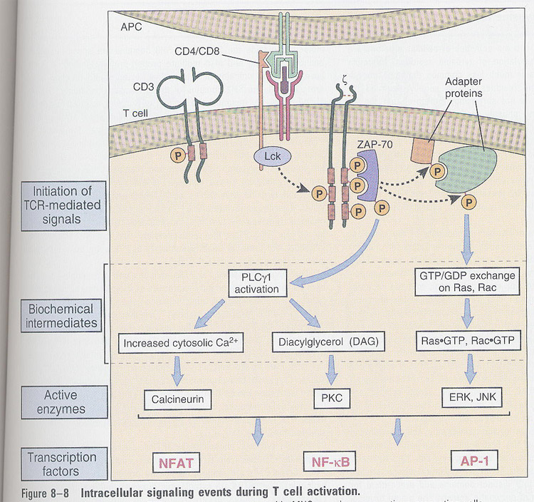
** T cell activation (Fig 9-8)
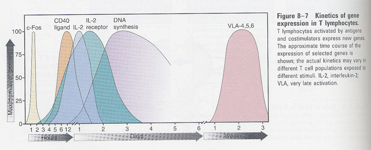
- steps:
1. early signal
transduction
2. transcriptional
activation of a variety of genes
3. expression of
new cell surface molecules
4. secretion of
cytokines (ex; IL-2)
5. induction of
mitotic activity
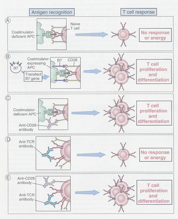
- two signal models
1. by Ag-MHC and
TCR
2. costimulation
by accessory molecules (ex; CD28): (Fig 9-6)
** Role of costimulators in T cell activation
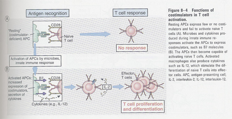 Fig
9-6
Fig
9-6
Ref; TCR and accessory molecules
Fig/Fig6-1.jpg)
---> Role of B7 and CD28 in T cell activation
.jpg)
-- Role of CD40 in T cell activation
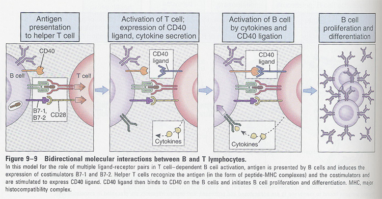 Fig 10-11
-- T cell signal transduction
 Fig
9-9
Fig
9-9
(1) tyrosine phosphorylation
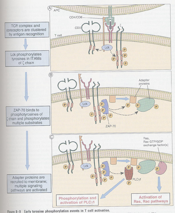 Fig
9-10
Fig
9-10
(2) Ras/MAP kinase pathway
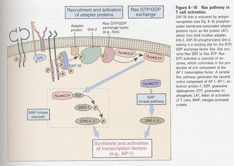 Fig
9-12dmd
Fig
9-12dmd
(3) inositol lipid metabolism
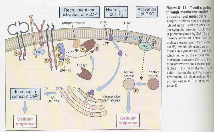 Fig 9-13
(4) Activation of transcription factors in T cells
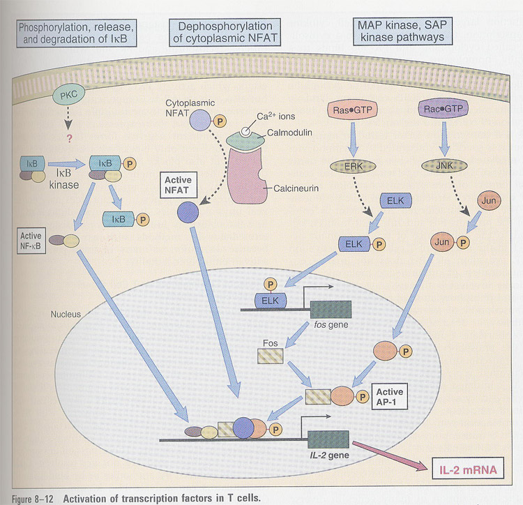 Fig 9-14
Chapter 10 B cell activation and antibody production
General features of humoral immune response
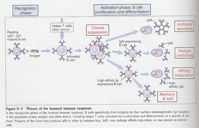 Fig 10-1
(a) primary and secondary
antibody response
- cell cycle from G0 to G1
- proliferation
- Ig secretion
- isotype switching
(b) intracellular signal changes
- Ca2+, DAG, PKC activation (within minutes)
- c-fos and c-myc activation (within 30'-1hr)
- size and cellular RNA increase (~12hr)
- cytokine receptors and class II expression (within
12-24hr)
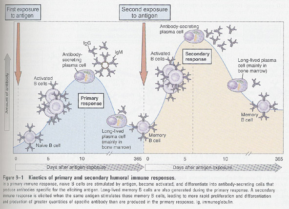 Fig
10-2
Fig
10-2
Humoral immunity:
mediated by antibodies
1. Antibody responses: Ag + resting
B, IgM/IgD mature cells
--
proliferation (growth)
--
differentiation (Abs secretion)
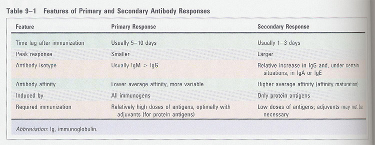
2. Features of antibody responses:
(a) presence of T cells; two signals (one from Ags, the other from
T cells and their products)
(b) in case of non-protein antigens; no need T cells
(c) different primary and secondary responses: (Fig 10-2)
___________________________________________________________
primary secondary
___________________________________________________________
10,000-1,000,000 fold 1-100ng/ml
of specific Ags
macrophages/dendritic cells high
affinity Igs on B cells
(non-specific) (only
in T-dependent Ags)
____________________________________________________________
(d) memory cell generation/heavy chain class switching/affinity
maturation
Signal transduction by the BCR complex
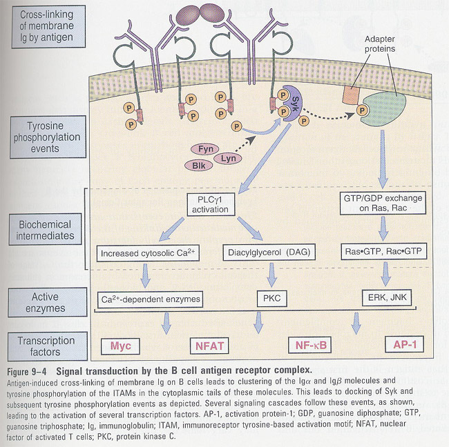 Fig 10-4
Second signals for B cells provided by complement receptors
Fig/Fig9-5.jpg) Fig 10-5
Fig 10-5
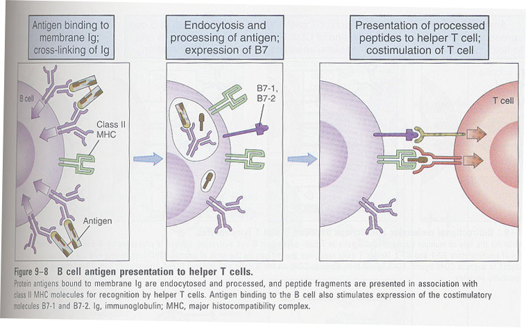
Functional response of B cells to antigen recognition
Fig/Fig9-6.jpg) Fig 10-6
Fig 10-6
Which effects are coming up when Ags are introduced into B cells ?
(1) 2 types of responses
:
--
entry of resting B cells into the cell cycle
--
antigen processing
(2) functional aspects: -- phosphorylase
C catabolization
--
c-fos and c-myc
--
size and cellular RNA increase
--
cytokine receptor/MHCII
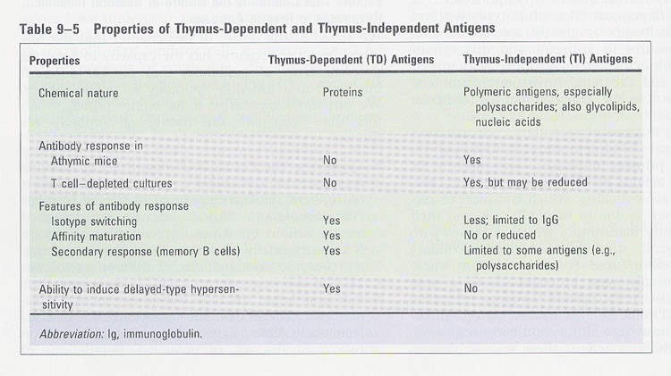
Antigens
(a) thymus dependent and independent Ags
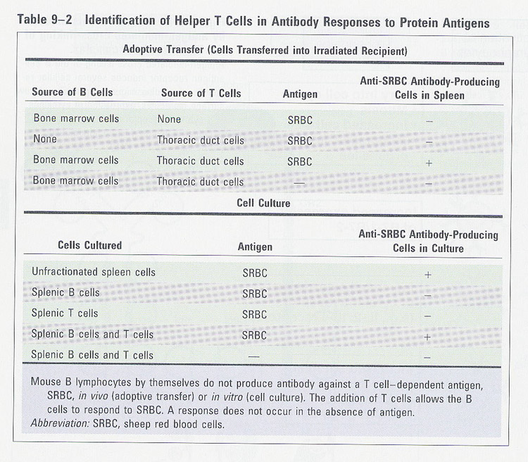
B-T cell interactions
(a) How are T helper cells found
to be involved in B cell activation ?
---> evidence
(b) early and late phase after immune response in lymph node
Fig/Fig9-7.jpg) Fig 10-7
Fig 10-7
참조 (Germinal Center): Fig 10-12
(c) B and T cell migration and interaction
Fig/Fig9-8.jpg) Fig 10-8
Fig 10-8
(d) How B cells interact with T cells for immune response ?
Fig/Fig9-9.jpg) Fig 10-9
Fig 10-9
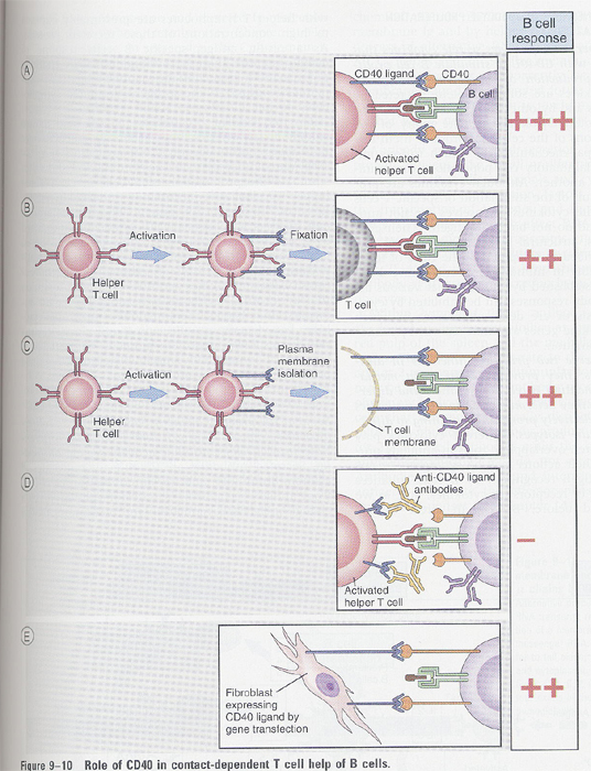
Role of CD40 in B cell activation
Fig/Fig9-10.jpg) Fig 10-11
Fig 10-11
--> experimental evidence
Secretion of Ig
- activation of mIg expressing B cells by antigen, in plasma cells
- more differentiation, more sIg
- mRNA splicing by choice of poly A sites
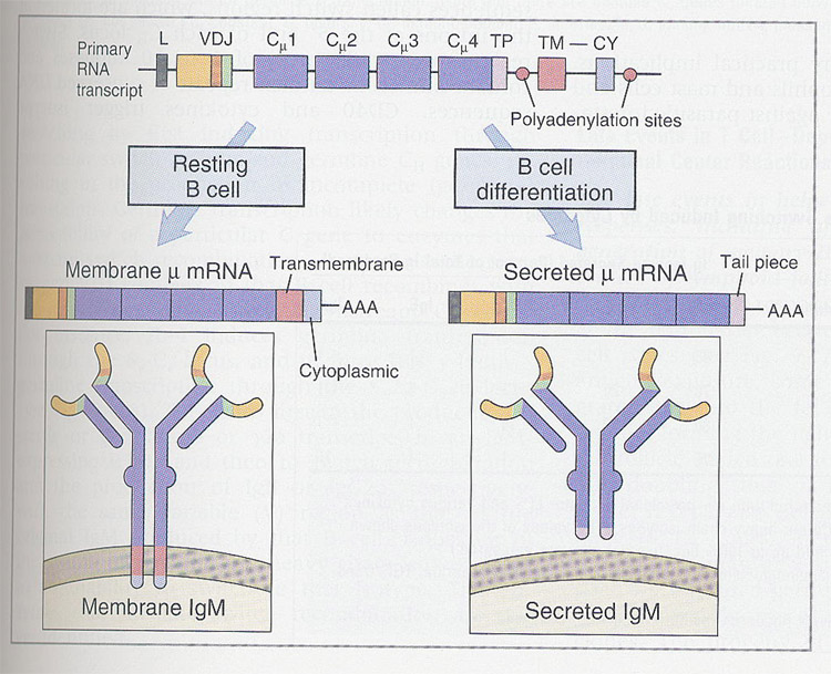 Fig 10-18
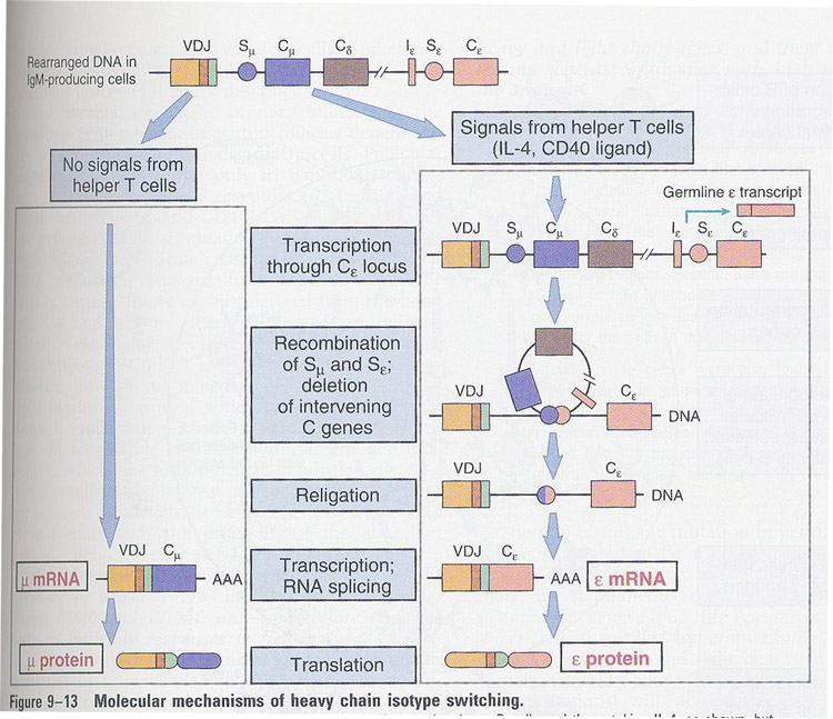
H chain class switching
- in IgM/IgD B cells by antigen
stimulation
- mechanism: (Fig
10-14) -- switch recombination
switch
region (5' each C region), ex; for IgE
-
1-10 kb long, numerous tandom repeat of conserved seq (52bp)
- regulation: 1) by helper T cell and its cytokines
2)
by microenvironment
-
opening the chromatin, and accessible to factors regulating rearrangement and
transcription in switch region
-
sterile C transcript (not VDJ) in short time after treatment
examples:
IL-4 (IgE), gamma-IFN (IgG2a), mucosal tissues (IgA)
 Fig
10-14
Fig
10-14
Fig/Fig9-13.jpg) Fig 10-13
Fig 10-13
Germinal Center Reaction: Late event in immune response
Fig/Fig9-15.jpg) Fig 10-12
Fig 10-12
- Somatic mutations of Ig in germinal center
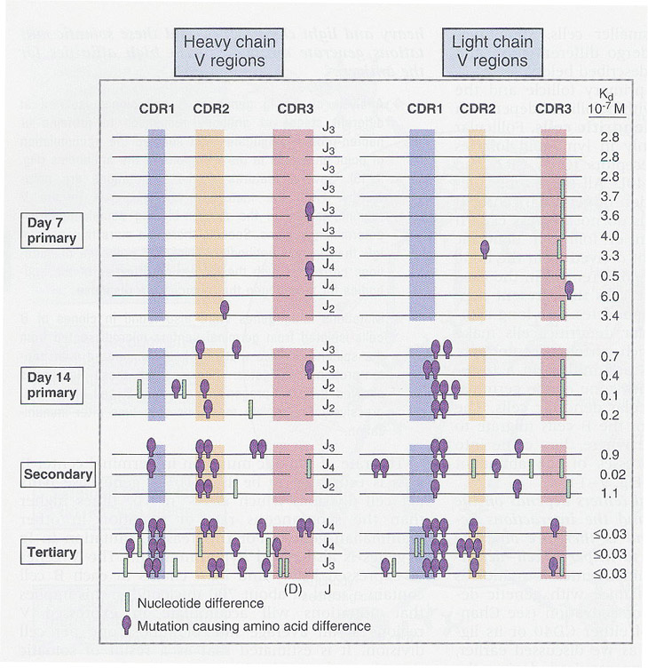 Fig
10-16
Fig
10-16
1. in antigen stimulation, occurrence in V (H and L) region, affinity
maturation
2. characteristics;
- number of mutations increase in IgG>IgM anibody, 3'>2' immunization,
and with time after 1' immunization
- hypervariable region of V exon is frequent (affinity maturation)
- some mutations: loss of Ag binding activity - die
other mutations: new Ag specificity
- 1/1000 per base pair in V region
- affinity maturation: in antibody response to helper T cell - dependent
antigens
T cell or T cell derived products
B cell selection in Germinal Center
Fig/Fig9-18.jpg) Fig 10-17
Fig 10-17
Antibody feedback
definition: injection of Ag-specific
antibody cause the reduction of Abs production
in both humoral
and cell-mediated response
(ex: Rh disease)
---> ♂(Rh+) ♀(Rh-)
How ? --> SHIP(SH2 domain containing inositol phosphatase) --> removal of PIP3
--> no signaling by BCR
Fig/Fig9-19.jpg) Fig 10-19
Fig 10-19